5 MIN READ
Corn Leaf Diseases
January 30, 2023
Key Points
- Different diseases may have similar symptoms, particularly during the early stages of disease development.
- It is not uncommon for a corn plant to have several different diseases present at the same time.
- Multiple diseases present on a corn plant can make disease diagnosis difficult.
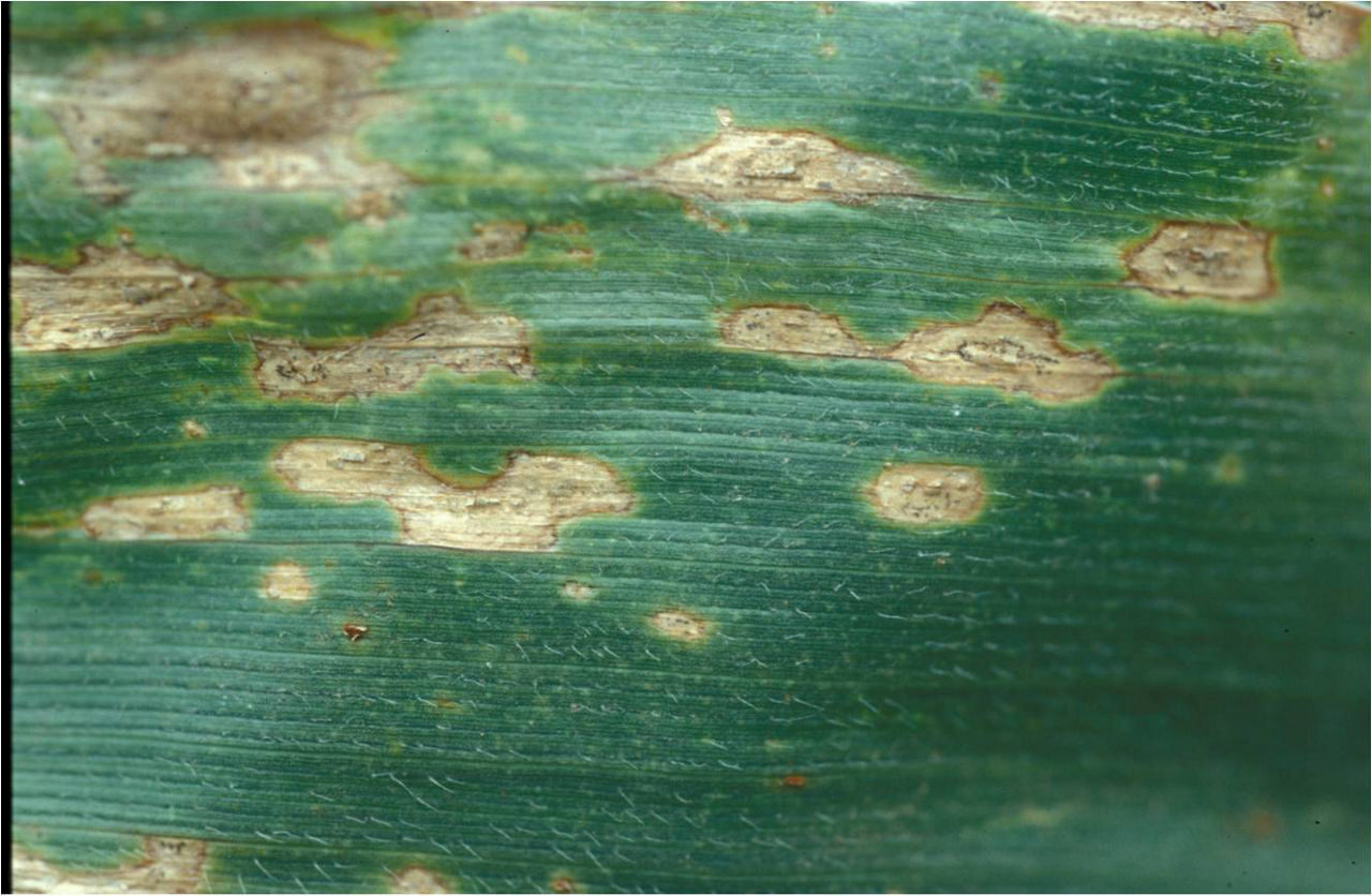
Anthracnose Leaf Blight
Symptoms of anthracnose leaf blight include small, oval to elongated, water-soaked lesions that appear on youngest leaves and turn tan to brown with yellow to reddish brown borders (Figure 1). Small, black, hair-like structures (called setae) may sometimes be seen in the middle of lesions. Lesions may coalesce, blighting the entire leaf. Heavily infected leaves wither and die. Leaf symptoms are most common on the lower leaves early in the season and on the upper leaves late in the season. Infection occurs in warm, humid weather. The same fungal pathogen is responsible for both anthracnose leaf blight and stalk rot; however, the presence of leaf blight does not necessarily indicate that stalk rot will be a problem later in the season. The stalk rot phase is of greater concern than the leaf blight phase.
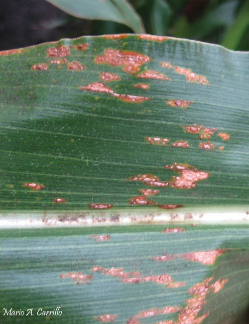
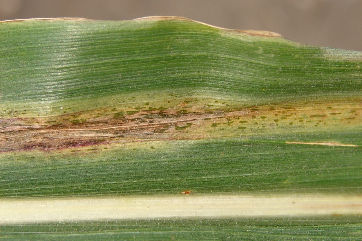
Common Rust
Common rust causes small, cinnamon-brown, powdery, circular-to-elongated pustules to occur on upper and lower leaf surfaces, often in bands across leaves (Figure 2). In contrast, pustules of southern corn rust are orange-colored and occur primarily on the upper leaf surface. Rust pustules rupture the leaf surface (epidermis) and powdery rust spores can be rubbed off. Pustules become dark brown to black late in the growing season. The disease is favored by moderate to cool temperatures and high humidity. The fungus does not overwinter in the Corn Belt.
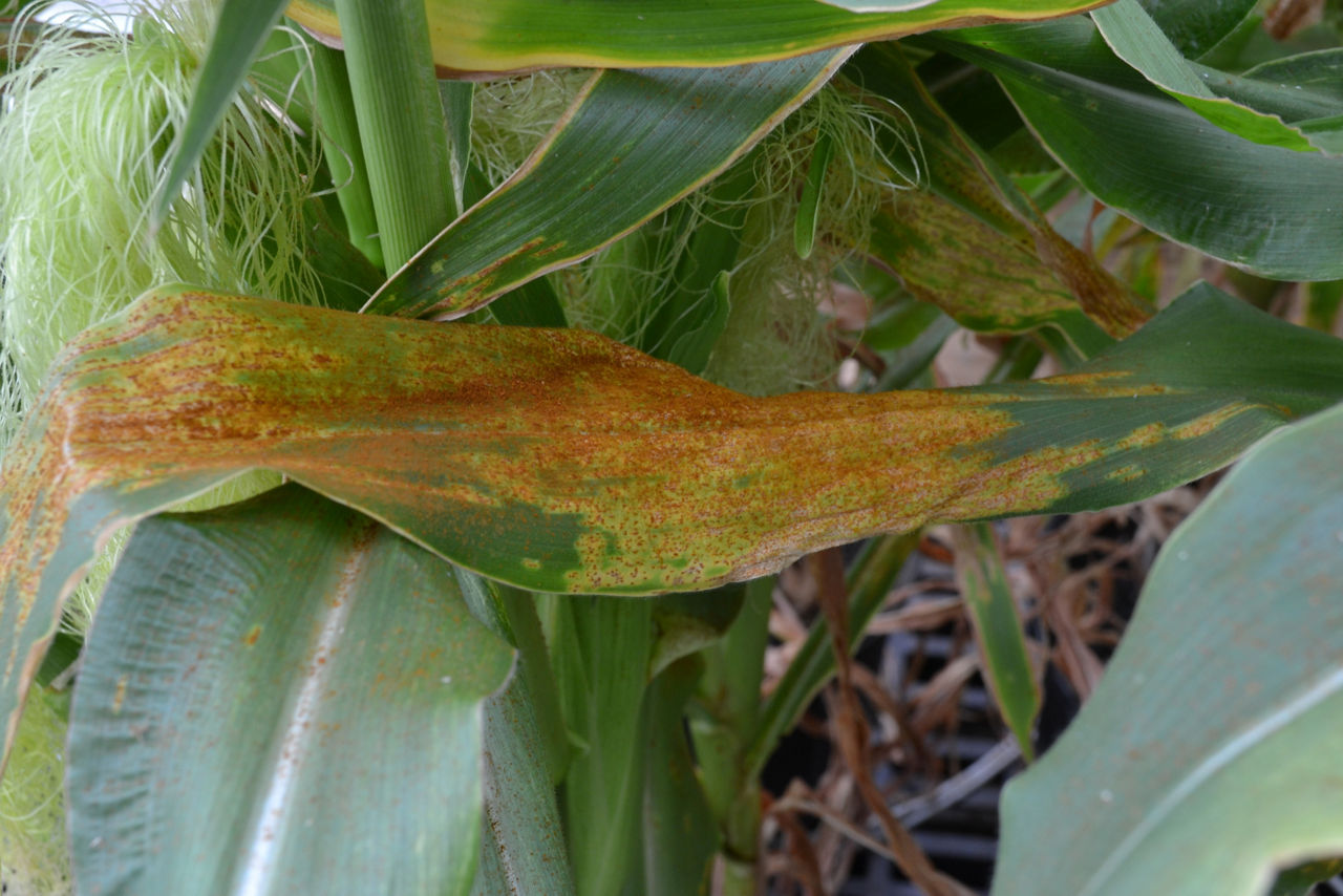
Southern Rust
Similar to common rust, southern rust symptoms are small, circular, light cinnamon-brown to orange-colored pustules occur on upper surfaces, leaf sheaths, and husk leaves (Figure 3). Pustules often are very dense in areas of infected tissues. Pustules break the leaf surface (epidermis) less frequently than common rust. Infection is favored by warm, humid weather.
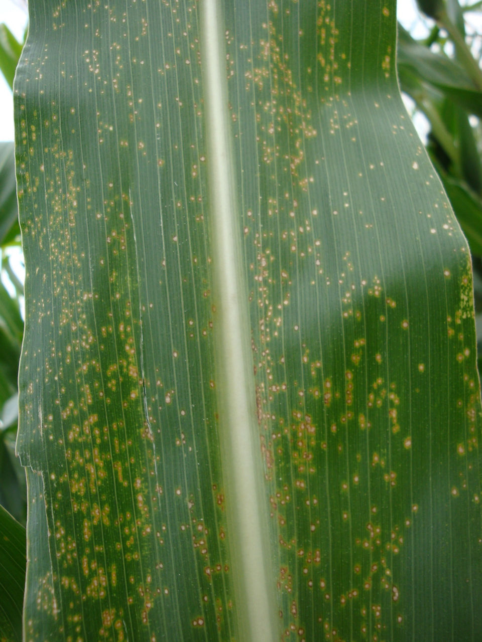
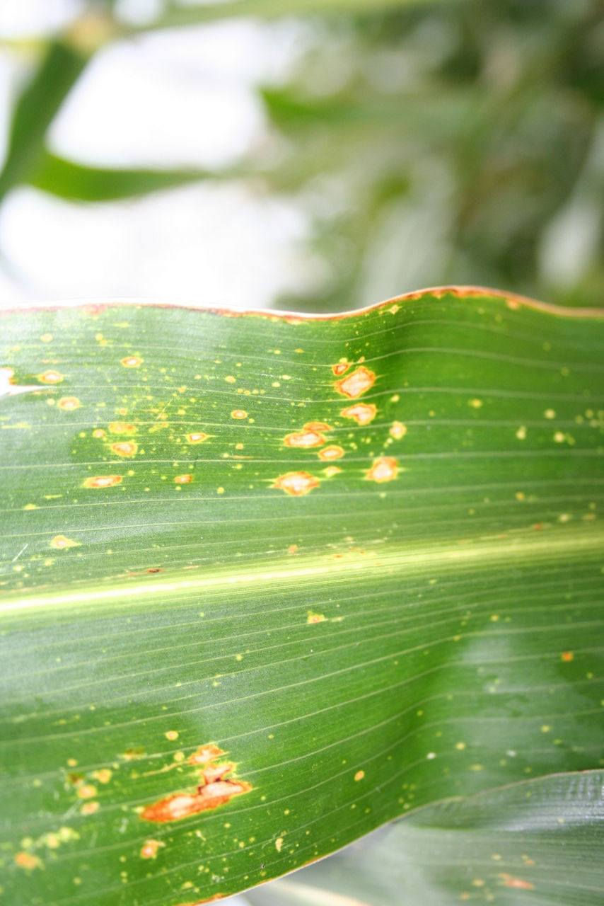
Eyespot
Eyespot appears as small, circular to oval, translucent lesions surrounded by yellow to purple margins that gives them a halo effect (Figure 4). Lesions occur on leaves (most commonly as plants approach maturity), sheaths, and husks. The disease is favored by cool, moist weather.
Gray Leaf Spot
Gray leaf spot has gray to tan, rectangular lesions on leaf, sheath, or husk tissue (Figure 5). Spots are opaque and long (up to 2 inches). Lower leaves are affected first, usually not until after silking. Lesions may have a gray, downy appearance on the underside of leaves where the fungus sporulates. The organism thrives in extended periods of warm, overcast days and high humidity. Gray leaf spot has become more prevalent with increased use of reduced tillage and continuous corn.
Physoderma Brown Spot and Node Breakage
Physoderma brown spot first has small yellow spots that appear at the base of the leaf and over time turn brown in color. As infection progresses, spots can often be found occurring in bands across the leaf. Spots in the mid-rib of the leaf become reddish to brown in color and combine to form irregular blotches (Figure 6). Sheath, husk, tassel, stalk, and leaves may exhibit symptoms late in the season. Infected stalks may break at a node. This disease is favored by warm, wet weather.
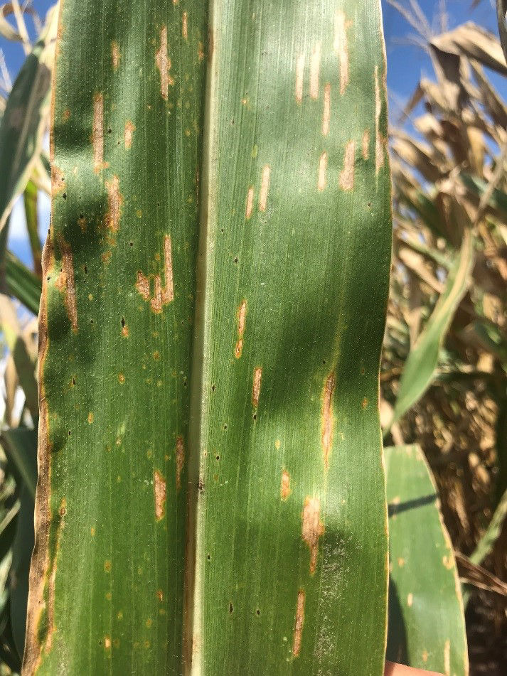
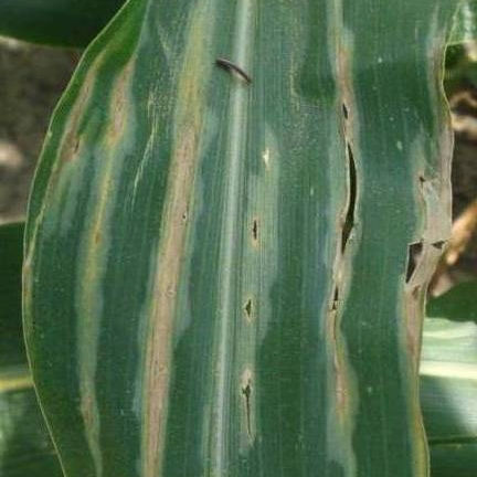
Northern Corn Leaf Blight
Long (up to 6 inches), elliptical to cigar-shaped, gray-green lesions that become tan, brown are symptomatic of infection by northern corn leaf blight (Figure 7). Infection begins on lower leaves and moves up the plant. Lesions may form in bands across leaves because of infection in the whorl. The disease is favored by high humidity and moderate temperatures.
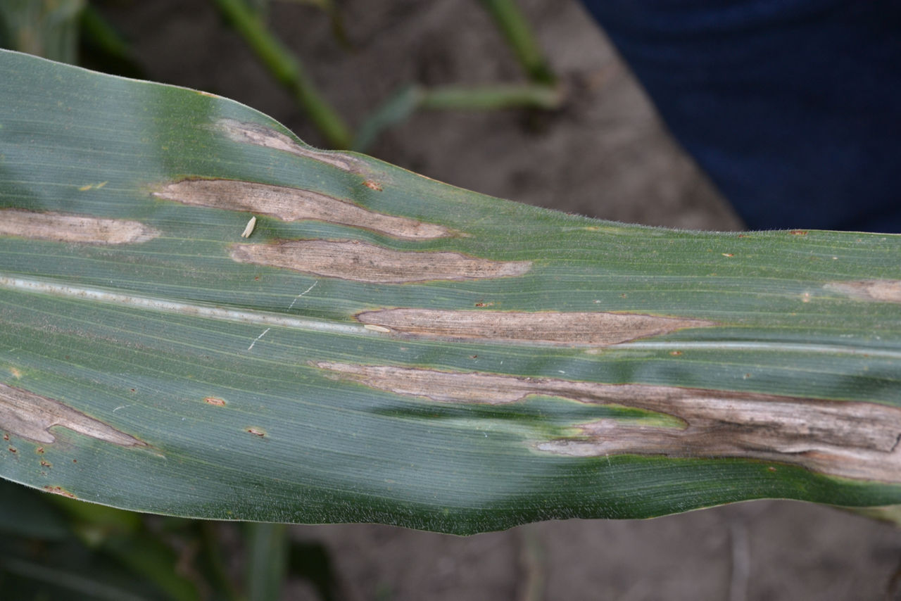
Southern Corn Leaf Blight
Southern corn leaf blight produces small, elongated (up to 1-inch long) parallel-sided lesions that are tan with brownish borders (Figure 8). Symptoms vary considerably on different corn products, often requiring microscopic examination of the fungal structures to confirm diagnoses. This blight primarily attacks leaves and is favored by high humidity and warm temperatures.
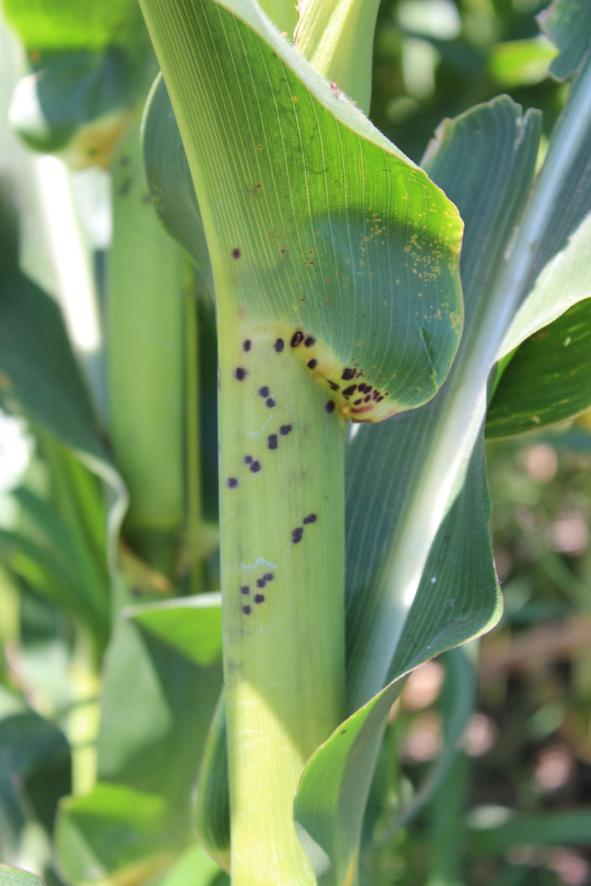
Goss’s Wilt
This bacterial disease causes both a seedling and adult-plant wilt. Adult plant wilt is typically associated with leaf blight, not Goss’s Wilt. Systemically infected seedlings may wilt and die. Vascular bundles can be discolored. More common later-season infections of leaves produce dull gray green to necrotic lesions often with irregular margins. Small, water-soaked “freckles” appear within developing lesions (Figure 9). Bacterial droplets may ooze from infected tissues early in the morning, leaving a shellac-like appearance when dried on leaf surfaces. Plant injury, such as hail or wind damage, enhances infection.
Stewart’s Bacterial Wilt
Symptoms of Stewart’s wilt or Stewart's disease on leaves are long, green-gray, water-soaked lesions with wavy margins, accompanied by stunting and wilting which may lead to plant death at the seedling stage (Figure 10). Cavities may form in the stalk near the soil line. The more common leaf blight phase appears after tasseling. Leaves are streaked with gray green to yellow-green lesions, each distinguished by the presence of a flea beetle feeding scar toward the base of the streak. Streaks are long and irregular, turning tan as the tissue dies. Flea beetles are the primary vector, and incidence of the disease is relative to the size of the beetle population.
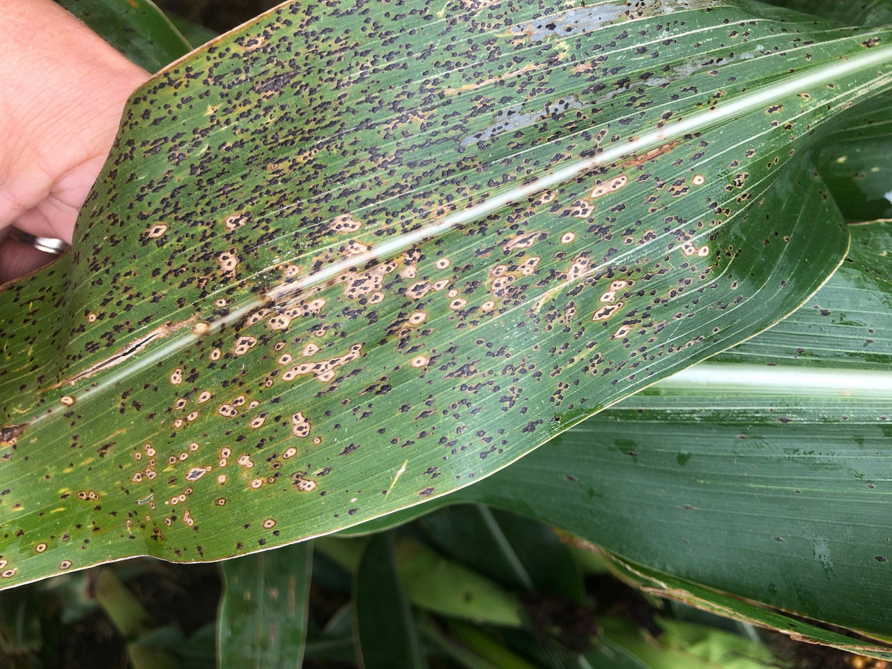
Tar Spot
A relatively new disease, tar spot was first found in the Midwest in the mid 2010’s. It is a fungal disease and is primarily found on the leaves. The symptoms include small irregularly shaped black lesions on both upper and lower leaf surface that cannot be rubbed off (Figure 11). It can also produce lesions that have a halo appearance with the black spots surrounded by a tan to yellowish halo circled with a dark border, referred to as “fish-eye” lesions. A field diagnostic technique is to wet the leaf and rub the lesions between your thumb and index fingers -- tar spot lesions will not rub like rust spores on the leaf surface, for example. Corn products that are susceptible can be infected at any developmental stage when conditions are favorable. The pathogen overwinters on infested corn residue and serves as a source of inoculum for the following season. Management includes burying residue, crop rotation, tolerant corn products, and fungicides.
Management
Timely scouting is important to help protect corn plants from diseases caused by fungal pathogens. Since much of a corn plant’s energy from photosynthesis is produced by the leaves immediately surrounding the primary ear, those leaves should be protected from foliar diseases, if possible. Fungicide applications made before a fungal disease spread throughout the corn canopy may help maximize yield potential in high disease pressure environments. However, since Goss’s Wilt and Stewart’s wilt are both caused by bacterial pathogens, these diseases are not affected by fungicide applications.
Fields with foliar diseases should be scouted for stalk health as the reduction in photosynthesis can predispose corn plants to stalk lodging. Identification of foliar diseases can help determine the need for future management practices such as tillage, crop rotation and the selection of disease-tolerant corn products to help reduce disease occurrence next season.
Sources:
1 Bissonnette, S.M., Pataky, N.R., Nafziger, E.D., et al. 2010. Field crop scouting manual. X880e. University of Illinois Extension.
2 Brouder, S.M., Camberato, J.J., Casteel, S.N, et al. 2014. Corn and soybean field guide, 2014 edition. ID-179. Purdue University.
3 White, D.G. 1999. Compendium of Corn Diseases, third edition. The American Phytopathological Society.
4 Hershman, D.E., Vincelli, P., and Kaiser, C.A. 2011. Foliar fungicide use in corn and soybean. University of Kentucky. PPFS-GEN-12. http://plantpathology.ca.uky.edu/.
5 Kleczewski, N. et al. 2019. Tar spot. Crop Protection Network. https://crop-protection-network.s3.amazonaws.com/publications/tar-spot-filename-2019-03-25-120313.pdf
1211_61471
Disclaimer
ALWAYS READ AND FOLLOW GRAIN MARKETING AND ALL OTHER STEWARDSHIP PRACTICES AND PESTICIDE LABEL DIRECTIONS.